Evaluation of pericardial catheter placement versus needle pericardiocentesis in the dog | VETgirl Veterinary Continuing Education Podcasts


In today’s VETgirl online veterinary CE podcast, we will be evaluating two methods of pericardiocentesis based off a study by Cook et al entitled Prospective evaluation of pericardial catheter placement versus needle pericardiocentesis in the management of canine pericardial effusion. Pericardial effusion in dogs is a condition we see with some degree of frequency in the ER. The urgency of this condition arises from the buildup of trapped fluid in the small sac surrounding the heart, located between the pericardial lining and the myocardium. Normally this space is so small that you can’t really see it on an emergency ultrasound scan without a cardiologist and a diagnostic ultrasound. When enough fluid builds up in the pericardial sac, this space becomes easier to see on emergency ultrasounds. But more importantly, as the fluid accumulates, the pressure in this trapped space compresses the heart chambers, preventing adequate filling of the heart, and results in less blood exiting the heart, and so less blood and oxygen is supplied to our vital organs. This condition is called cardiac tamponade and represents one of the causes of “obstructive shock.” Clinical signs that may clue us in to the presence of cardiac tamponade include Beck’s triad of: 1) low blood pressure 2) muffled heart sounds 3) and jugular venous distention, and often the patient is also tachycardic which is the body’s compensatory response to the lower cardiac filling volumes.
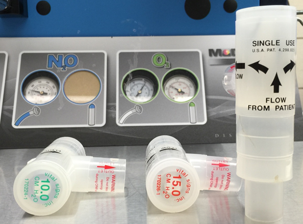
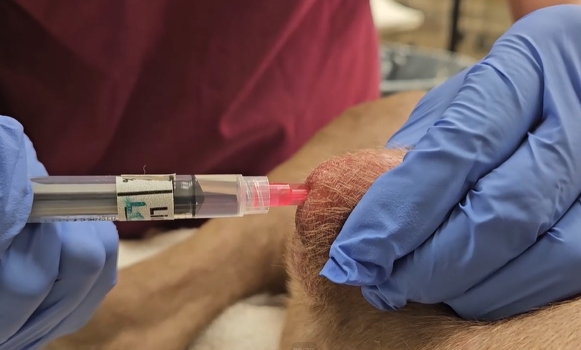
Pericardial effusions may develop from idiopathic or neoplastic processes (which are the most common causes in the dog), or can result from infection, coagulopathies, atrial rupture, severely hypoproteinemia, PPDH, and congestive heart failure. Urgent treatment of cardiac tamponade requires the expedient drainage of the fluid from the pericardial sac to relieve pressures on the cardiac chambers. Often a needle or venous catheter is used for percutaneous insertion through a fast-prepped, right-sided thoracic site (either side may be used, but right side has an exaggerated cardiac notch between lung lobes), and advanced into the pericardial space under slight negative pressure until fluid is retrieved, or advanced under ultrasound guidance until insertion into the pericardial space is identified. You can always check out our VETgirl Real-Life Rounds on how to perform a pericardiocentesis [January 4, 2018: VETgirl Real-Life-Rounds: How I (quickly) approach pericardial effusion] or watch the VETgirl video on how to perform a pericardiocentesis..
At the time of initial pericardiocentesis, clinicians are often unaware of the etiology of the pericardial effusion, and therefore are unable to make an educated prediction on whether the fluid will build up again and necessitate another drainage, and how fast this may occur. A patient could theoretically require multiple pericardial drainage procedures within the same hospital stay, or may be discharged and never require another. In humans, it is common practice to place a pericardial catheter to facilitate repeated elective drainage every 4-6 hours; this is referred to as “extended pericardial catheter drainage” or EPCD for short. The benefits of a pericardial catheter include ease of repeated drainage and instillation of medications such as sclerosing agents or chemotherapy agents directly into the pericardial space. Our main veterinary use for pericardial catheter placement is mostly to facilitate the need for frequent drainage of pericardial effusion, especially in conditions that may produce large volumes of pericardial effusion such as cardiac neoplasia. A 2019 retrospective study on pericardial catheter placement in dogs showed that percutaneous placement of chest tube catheters by the modified Seldinger technique was generally well tolerated under light anesthesia and carried only a mildly increased risk of arrhythmias (22%) as compared to using a needle alone for single-use drainage (13%).(1) These same authors then created this current prospective study, Cook et al (that we will refer to for the remainder of the podcast), and it was meant to evaluate the practicality of placing these catheters in the clinical setting and to assess whether the presence of these catheters over the course of days may affect the risk for arrhythmias, and finally to assess the general recurrence of pericardial effusion among their patient population (2). An unfortunate and frequent occurrence in clinical veterinary studies is that they are often underpowered because of the difficulty in obtaining client consent or collecting enough subjects that meet the study’s inclusion criteria. This particular study was able to recruit 31 dogs, but the authors make mention that 48 was the number they were striving for.
Causes of canine pericardial effusion in this study matched those of prior studies supporting neoplasia and idiopathic as the top two causes in dogs. 31 dogs that presented to the teaching hospital and were subsequently diagnosed with pericardial effusion were randomized to either a needle pericardiocentesis group or a pericardial catheter placement group. There was no significant difference in age or gender between the groups, though the needle group consisted of more intact dogs. Most dogs were large breeds such as Labs, German Shepherds, Mastiffs, Boxers, and others. Needle pericardiocentesis was achieved by percutaneous insertion of large bore peripheral catheter stylettes attached to extension tubing. For the pericardial catheter group, clinicians used 20cm chest tubes inserted percutaneously by modified Seldinger technique, with or without ultrasound guidance. Light sedation for the procedures was achieved mainly with midazolam and frequently accompanied by butorphanol, but selection was at the clinician’s discretion. One brachycephalic dog was fully anesthetized for better airway control owing to the inherent risk presented by this breed’s respiratory anatomy. Out of 15 patients assigned to the pericardial catheter group, 3 catheters were unable to be placed, 9 were successfully placed on first attempt, and 3 were successfully placed on second attempt. Since this study did not require the use of ultrasound-guidance to assist in catheter placement, it would be interesting to see if success rates for pericardial catheter placement could be improved by use of ultrasound-guidance.
Catheters were left in place for 14-85 hours and drained at the clinician’s discretion every 4-6 hours, so there was no standard drainage protocol nor standard catheter duration. One dog in the pericardial catheter group required urgent drainage 12 hours after catheter placement to remove 10.6 ml/kg of fluid produced by a right auricular mass. 4 cases had negligible fluid drainage of <1 mL/kg total, but the authors did not state whether clinicians proved these catheters were patent; one such way could have been to document on ultrasound the lack of visible pericardial effusion to suggest the catheters were effectively draining. 3 cases had single event drainages of 8.5-10.6 ml/kg within 12 hours after catheter placement. Overall, the median pericardial fluid production of the 12 dogs was 0.09 ml/kg/hr over 21 hours total, with a range of 0 to 0.76 ml/kg/hr. Catheters were removed due to euthanasia in 5 of the 12 patients, at time of discharge in 3 of the 12, and the rest were pulled at undisclosed times and at the preference of the attending clinician. One of the main goals of this study was to determine if placement of pericardial catheters is practical enough to perform at time of initial pericardial drainage. This study suggests the time from catheter insertion to achieving effective drainage is similar enough to the time spent when using a needle for pericardial drainage, and so we can consider placing pericardial catheters at the first emergency drainage event. According to the authors, the most time-consuming step of the pericardial catheter placement is securing the catheter to the patient, but note that once the catheter is within the pericardial space, drainage can commence immediately while the catheter is being sutured and wrapped in place; so this is not lost time. The second main goal of this paper was to determine if pericardial catheters resulted in greater arrhythmia formation compared with pericardial drainage through needle insertion. Of the dogs that presented with sinus rhythms, 4 of 11 dogs in the pericardial catheter group experienced arrhythmias during catheter placement compared with 9 of 14 dogs in the needle pericardiocentesis group. The most common types of arrythmias encountered were frequent VPC’s and ventricular tachycardia (defined as sustained ventricular complexes >30s at a rate >160 bpm). Other arrhythmias identified included APC’s, second degree AV block, and accelerated idioventricular rhythm. It was determined that there was no significant difference in arrhythmia formation during or after the procedure between groups. This means that, according to this study, the insertion and presence of a pericardial catheter doesn’t appear to be more arrhythmogenic than traditional methods of pericardial drainage. However, we must review this statement carefully as the true incidence of arrhythmias may have been underreported due to the method of ECG monitoring implemented in this study. ECG’s were recorded in the form of 2-minute spot-checks every 4 hours instead of continuous ECG monitoring, which may have resulted in under-reporting of arrhythmias. But we can say, from a clinical perspective, that no life-threatening arrhythmias were detected as a result of the pericardial catheter. To better explore the safety of these catheters, follow-up studies utilizing continuous ECG throughout hospitalization coupled with a consistent duration of pericardial catheter placement may prove helpful.
The last goal of this study was to determine if pericardial catheters could prevent our patients from requiring repeated PE drainage and assess recurrence rate of PE over time. Within the pericardial catheter group, one patient did require emergency drainage of fluid, but it remains unclear if others would have required an emergency drainage since all catheters were periodically drained. Within the needle group, 20% of patients required repeated drainage within 24 hours, so perhaps this provides more telling information of the recurrence rate of PE’s. The recurrence of PE overtime couldn’t be assessed in this study as many dogs were euthanized (assumed due to the poor prognosis of their disease), or they received surgical intervention for their disease, or they were lost to follow-up.
So what can we take away from this VETgirl podcast? It appears that pericardial catheter placement in dogs can be performed in a time-efficient manner on an emergency-basis if needed. We can perhaps say from this study that pericardial catheter placement does not carry a greater risk for arrhythmia generation, but more studies are needed to explore whether the continued placement of a pericardial catheter can generate arrhythmias over time. Based on this study, pericardial catheters do not appear to carry a high risk of causing life-threatening arrhythmias when left in place for days. The clinical usefulness of these catheters should be considered against the ultimate goals for the pet and their family. If it is determined that the pet needs a definitive surgical intervention, such as a pericardial window, perhaps a pericardial catheter can save ER staff time and resources and provide comfort for the pet as opposed to repeated needle pericardiocentesis. With such a low reported 24 hr recurrence of PE at 20%, perhaps it’s not warranted to recommend the placement of pericardial catheters in all cases of PE at this time. Future studies are needed to fully evaluate the risk of post-placement arrhythmias, and to investigate on an individual PE-producing disease basis what the cost or health benefits of pericardial catheter placement are over traditional pericardial drainage before they can be recommended as a mainline treatment for specific diseases.
References:
1. Cook S, Cortellini S, Humm K. Retrospective evaluation of pericardial catheter placement in the management of pericardial effusion in dogs (2007-2015). J Vet Emerg Crit Care. 2019;29:413–417.
2. Cook S, Cortellini S, Humm K. Prospective evaluation of pericardial catheter placement versus needle pericardiocentesis in the management of canine pericardial effusion. J Vet Emerg Crit Care. 2021;31:11-17.
Today’s VETgirl online veterinary continuing education podcast is sponsored by IndeVets. IndeVets is creating a whole new way to work that puts vets like us first. They combine what makes associate work reliable with what makes relief work super flexible. That means all their doctors get proper pay for the hours they work, full benefits, and total schedule control – with a team dedicated to their happiness and growth every step of the way. IndeVets is vet-led and currently hiring highly competent and nice doctors. Check out them out at indevets.com – that’s I-N-D-E-V-E-T-S.com.
![]()
Not a VETgirl ELITE member? Get your learning on with the #1 veterinary CE resource – clinically relevant, practical, and accessible 24/7 on your own time! Don’t miss out – click HERE to check out your options, including our new 14-day, no-commitment ELITE Trial.


Only VETgirl members can leave comments. Sign In or Join VETgirl now!