


June 2026
In this VETgirl online veterinary continuing education blog, Amanda M. Shelby, RVT, VTS (Anesthesia & Analgesia) explores proper endotracheal tube (ETT) cuff inflation techniques in veterinary anesthesia. Discover how to avoid tracheal damage and overinflation risks in dogs, cats, and horses using evidence-based methods, like specialized cuff syringes, to protect patient airways and optimize anesthesia care.
How to Inflate an Endotracheal Tube Cuff to Minimize Risks, Maximize Benefit
By Amanda M. Shelby, RVT, VTS (Anesthesia & Analgesia), VETgirl Senior CE Specialist
Inflating the endotracheal tube (ETT) cuff is a routine step in the process of securing a patent airway following intubation. Surprisingly, 47.1-54% of veterinary professionals acknowledge that they inflate ETT cuffs in dogs, cats, farm animals, and horses based on how they were taught, while as little as 13.3-5.5% base their method of inflating the ETT cuff on peer-reviewed literature.1 Perhaps even more surprising, improper cuff inflation occurs as much as 86% in dogs.2 This seemingly small step to securing a patent airway is extremely important! In this VETgirl blog, we explore the various ways the ETT cuff is inflated in veterinary medicine and what can happen when it is over- and underinflated!
Murphy, or Magill, high-volume, low-pressure (HVLP) cuffed ETTs are the most commonly used ETTs in companion mammals (see Figure 1a). These are designed to distribute the cuff pressure over a larger surface area of the tracheal wall when creating a seal. Compared to low-volume, high-pressure cuffed ETT (see Figure 1b), modern HVLP ETTs are associated with lower inflation pressures to create a seal than LVHP ETTs.3 Additionally, they produce a lower incidence of tracheal irritation and complications; however, tracheal damage is possible when improperly overinflated.4-6
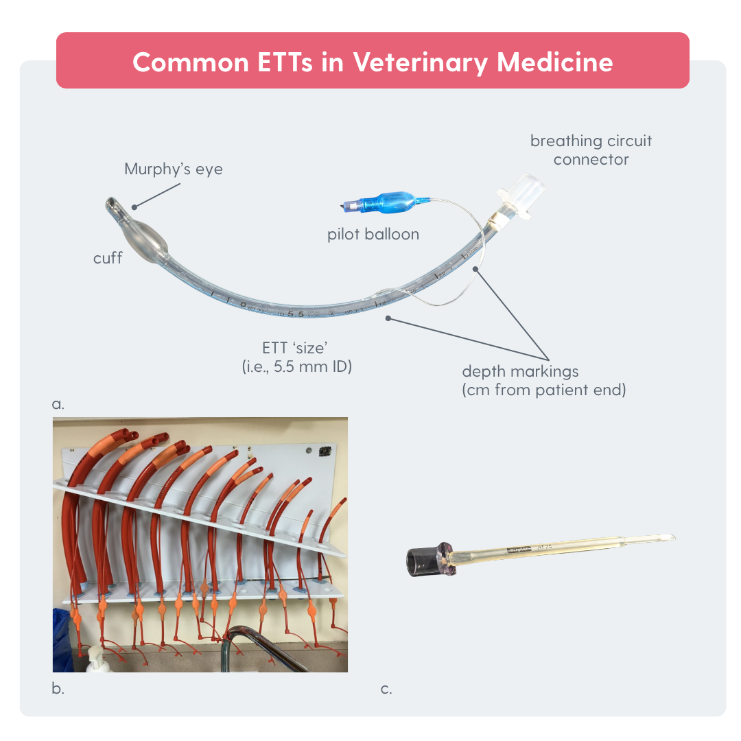
Figure 1: (a) High-volume, low-pressure, cuffed Murphy ETT (b) Low-volume, high-pressure cuffed ETTs (c) Cole ETT (Images courtesy of Amanda M. Shelby)
Regardless of which ETT is used, attention to proper cuff inflation and a thorough understanding of why we use ETT cuffs is important! When properly inflated, the seal produced by the ETT cuff reduces waste anesthetic gas (WAG) exposure and environmental pollution, assists in ensuring oxygen and inhalant delivery, reduces the risk of pulmonary aspiration, and helps facilitate ventilation and improved monitoring of ventilation with capnography. However, overinflation of the ETT cuff can result in tracheal irritation, tracheal mucosal wall ischemia, tracheal tears and resulting emphysema, tracheal stricture, and potentially increased airway resistance or obstruction from collapsing the endotracheal tube itself.5-9 While we don’t know the exact ETT cuff inflation pressure at which tracheal wall blood flow will be disrupted in cats and dogs, we generally follow the human guidelines of 20-30 cmH2O as being “safe”.10 Cuff insufflation pressure doesn’t always equal the pressure exerted on the tracheal wall – different ETT materials and sizes distribute pressure differently, and in hypotensive patients, blood flow may be disrupted at even lower cuff pressures (ugh, so much physics!).
When Do We Inflate the ETT Cuffs?
There are three distinct times we inflate ETT cuffs:
(1) During cleaning the tubes (if reusing your ETT—check out this VETgirl blog about best practices for cleaning and/or reusing ETT HERE)
(2) prior to use to ensure they are absent of leaks or tears, and
(3) following orotracheal intubation of the patient.
What are the Best Methods of ETT cuff Inflation?
Great news, several investigators have compared various methods for inflation of ETT cuffs. Generally, in small companion animals, the maximum peak inspiratory pressure (PIP) is 20-30 cmH2O, while in large animal species, these airway pressures are exceeded when conditions require various ventilation strategies with higher PIP.1 In short, our ETT cuff only needs to seal up to the point of the PIP as well as be sufficient to prevent aspiration of fluids.
Here are several commonly investigated methods for ETT cuff inflation in veterinary mammals:
Cuff Balloon Palpation:
This subjective method, likely carried over from the use of low-volume, low-pressure endotracheal tubes (see Figure 1b above) involves filling the ETT cuff pilot balloon until resistance or firmness is appreciated. Several investigators have demonstrated that ETT cuff balloon palpation frequently results in high intracuff pressures.11-12
Figure 2. ETT cuff balloon palpation (Video courtesy of Amanda M. Shelby)
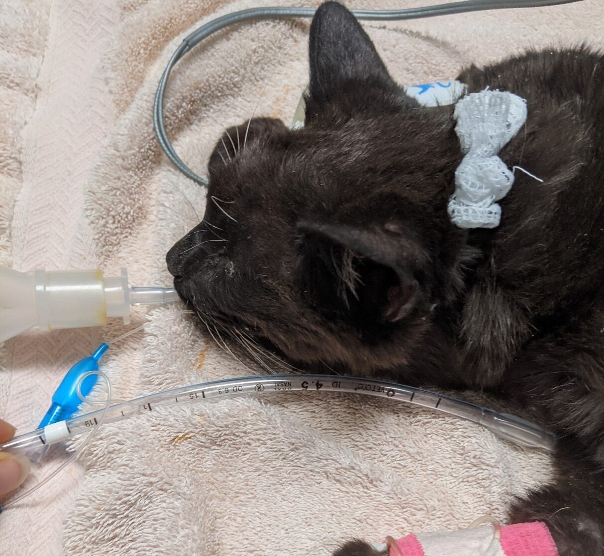
Minimum Occlusive Volume (MOV) (see Figure 3):
This method involves filling the ETT cuff with a regular syringe while delivering manual breath to the maximum peak inspiratory pressure (typically 20-30 cmH2O) while listening or feeling for a leak at the endotracheal tube. In a study that surveyed veterinary professionals’ methods for inflating ETT cuffs, this method was most common.8 But regardless of its popularity, this method has been shown to be effective at achieving optimal cuff inflation pressures compared to the use of specialized cuff inflation syringes.2, 12
Figure 3. ETT cuff inflation by MOV method (Video courtesy of Amanda M. Shelby)
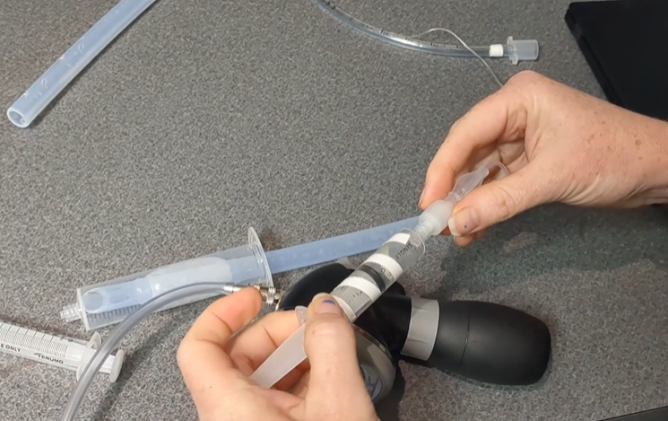
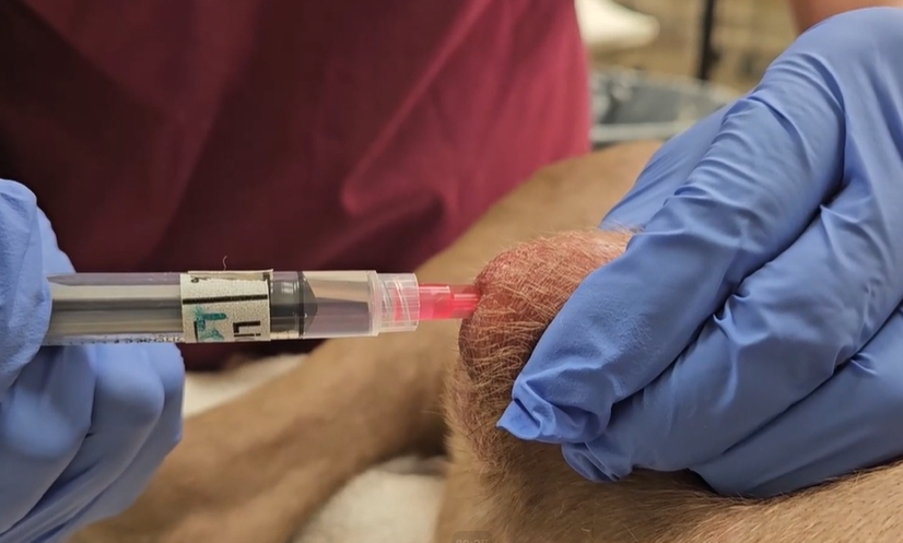
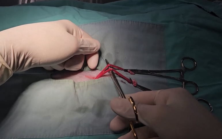
Loss of Resistance (LOR) with a Low Resistance Syringe (i.e., Perifix® LOR 10 ml; B-Braun or similar)(see Figure 4):
This method requires the anesthetist to initially overinflate the ETT cuff, then the low resistance syringe plunger is allowed to passively release excessive air from the cuff until a pressure equilibrium is established.
Figure 4. ETT cuff inflation by loss of resistance with a non-specialized syringe (Video courtesy of Amanda M. Shelby)
Specialized Cuff Inflation Syringe (see Figure 5 and 6):
This method involves using a commercially manufactured syringe or device that measures the pressure of the cuff or indicates when sufficient pressure is reached on the cuff that is associated with an ‘appropriate’ seal. Several brands have been validated and received FDA approval for use in humans (TruCuffTM [Anesthesia Equipment Supply, Inc. (AES)] and AG Cuffill® [Medline Industries, LP]).
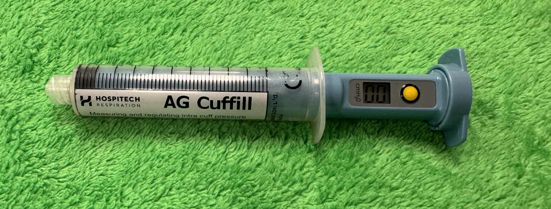
Figure 5. ETT cuff inflation device (Image courtesy of Joshua Madrid, RVT)

Figure 6. ETT cuff inflation device (Image courtesy of Jorgensen Laboratories, LLC. Product courtesy of Tru-CuffTM)
Observation of the Capnogram:
Some use the capnogram to evaluate the presence of a leak. In a leak-free anesthetic machine and patient breathing circuit, the ETT is the most likely source for a leak following induction of anesthesia and patient intubation. This can be seen as a decrease in the slope of the inspiratory portion of a capnogram (see Figure 7). Veen & de Grauw found this method to be the preferred method of ETT cuff inflation in horses.1 However, the study was a survey of current practices and did not provide ETT cuff pressure data.
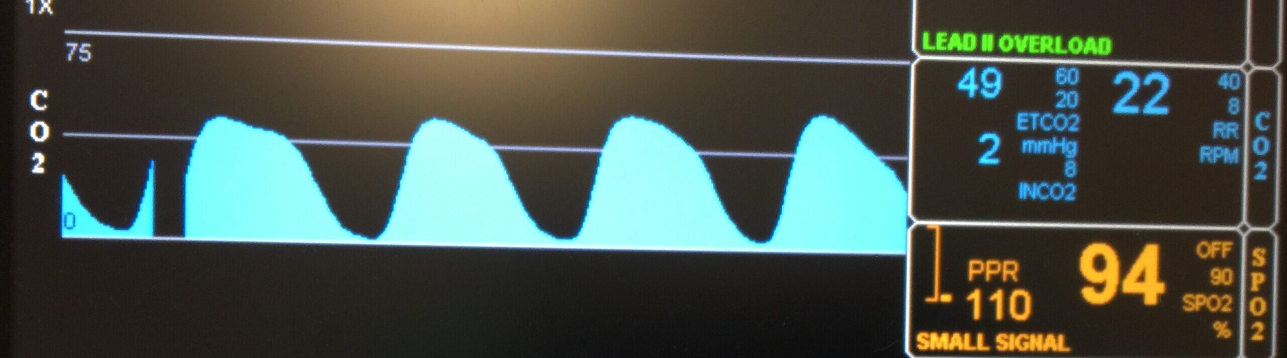
Figure 7. Capnogram in patient with a leak at the ETT (Photo courtesy of Amanda M. Shelby)
What Does the Literature Tell Us?
Findings from two studies are summarized in Figure 8.2, 12
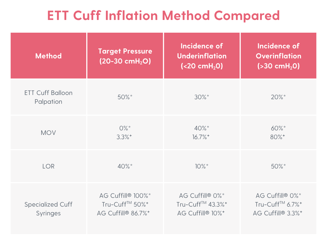
Figure 8: Incidence of various cuff inflation methods’ ability to accurately reflect target, under, or overinflation of the ETT cuff when represented by percentage (%) in various species. *Used a feline airway simulator,12 †Client owned dogs2
These studies found that the specialized cuff syringes produced the most reliable, targeted, intracuff inflation pressures between 20-30 cmH2O in dogs and in a study that used a feline airway simulator.2, 12
What About Large Animals?
ETT cuff inflation is just as important in these patients as well—perhaps even more so because ruminants salivate a LOT during anesthesia! Often in large animal patients, i.e., horses and cattle, following induction, we immediately place them on a ventilator, often disrupting the traditional MOV method of ETT cuff inflation. However, do not be discouraged…an additional palpation method involves ‘feeling’ for a leak by placing a hand on the ventral aspect of the patient’s ‘throat latch’ if it’s a horse or equivalent in a large ruminant during the delivery of a positive pressure breath. Some will use a stethoscope in this area to hear a leak. If a vibration is felt or heard, there is a leak! No vibration means no leak. Now, we could be overinflating or underinflating the cuff using this palpation method, and the use of a manufactured cuff inflation device could be superior, but to date, nobody has performed this study (wink wink, researchers!). Touzot-Jourde et al. found that ETT cuff pressures as high as 80-120 cmH2O in horses were required to effectively seal the airway during positive pressure ventilation with PIP of 30-40 cmH20.7 Additionally, these investigators concluded that at these cuff pressures, all ETT cuffs protected the airway from the aspiration of fluid they introduced but on gross and histologic examination of the tracheal mucosa, hyperemia, petechiae or hematomas and epithelial and submucosal damages occurred.7 Heath et al. found tracheal damage occurred in horses at ETT cuff pressures as low as 20 cmH20.9
Another common method of ETT cuff inflation in large animals, which I have personally experienced observing and taught to perform, includes inflating the cuff until resistance on a 30-60 mL syringe is experienced. This method is also likely to inflate the ETT cuff to excessive pressures, resulting in tracheal mucosal damage.
Why Didn’t We Talk About Non-Mammalian Species?
At VETgirl, we feel all living creatures are important! But many of our non-mammalian species use uncuffed or Cole ETTs. This is especially true for species that have complete tracheal rings (i.e., birds and reptiles). Sometimes uncuffed tubes are also used in pediatrics or small mammals prone to tracheal irritation and trauma.
VETgirl Key Takeaways:
• There currently are no evidence-based, veterinary-specific consensus guidelines on optimal cuff inflation practices or optimal pressure for veterinary species. However, of the studies available comparing ETT cuff inflation methods, digital syringes specifically manufactured for the purpose of inflating ETT cuffs provide the highest degree of accuracy for targeted ETT cuff pressures.
• Rarely are pressure manometers used to check ETT cuffs in veterinary practice.
• ETT cuffs may require frequent rechecking, inflation, or deflation to maintain an optimal cuff pressure.
• Using high-volume, low-pressure ETTs are recommended, but overinflation can still cause tracheal irritation and damage.
Abbreviations:
ETT endotracheal tube
HVLP high-volume, low-pressure
LOR loss of resistance
MOV minimal occlusion volume
PIP peak inspiratory pressure
WAG waste anesthetic gas
References:
1. Veen I & de Grauw JC. Methods Used for Endotracheal Tube Cuff Inflation and Pressure Verification in Veterinary Medicine: A Questionnaire on Current Practice. Animals (Basel). 2022;12(22):3076.
2. Hung WC, Ko JC, Weil AB, et al. Evaluation of endotracheal tube cuff pressure and the use of three cuff inflation syringe devices in dogs. Front Vet Sci. 2020;7:39.
3. Dorsch JA, Dorsch SE. Understanding Anesthesia Equipment. 5th ed. Philadelphia: Lippincott Williams & Wilkins; 2008. Chapter 19, p. 583.
4. Young PJ, Pakeerathan S, Blunt MC, et al. A low-volume, low-pressure tracheal tube cuff reduces pulmonary aspiration. Crit Care Med. 2006;34(3):632-9.
5. Loeser EA, Hodges M, Gliedman J et al. Tracheal Pathology Following Short-Term Intubation with Low-and High-Pressure Endotracheal Tube Cuffs. Anesthesia & Analgesia. 1978;57(5):577-579.
6. Seegobin RD, van Hasselt GL. Endotracheal cuff pressure and tracheal mucosal blood flow: endoscopic study of effects of four large volume cuffs. Br Med J (Clin Res Ed). 1984;288(6422):965-8.
7. Touzot-Jourde G, Stedman NL, Trim CM. The effects of two endotracheal tube cuff inflation pressures on liquid aspiration and tracheal wall damage in horses. Vet Anaesth Analg. 2005; 32:23–29.
8. Veen I, de Grauw JC. Endotracheal tube obstruction due to cuff overinflation or cuff herniation in small equids: A case series. Equine Veterinary Education. 2023;35(7):358-64.
9. Heath RB, Steffey EP, Thurmon JC, Wertz EM, Meagher DM, Hyyppa T, Van Slyke GL. Laryngotracheal lesions following routine orotracheal intubation in the horse. Equine Veterinary Journal. 1989;21(6):434-7.
10. Bird AR, Bird DJ, McMillan MW. Aspects of in vivo endotracheal tube intracuff pressure in cats. Vet Anaesth Analg. 2019;46(1):55-63.
11. Briganti A, Portela DA, Barsotti G, et al. Evaluation of the endotracheal tube cuff pressure resulting from four different methods of inflation in dogs. Vet Anaesth Analg. 2012;39(5):488-94.
12. White DM, Makara M, Martinez-Taboada F. Comparison of four inflation techniques on endotracheal tube cuff pressure using a feline airway simulator. J Feline Med Surg. 2020;22(7):641-647.
Only VETgirl members can leave comments. Sign In or Join VETgirl now!
It was interesting to read about the different methods of inflating ET tube cuffs. Some of these I had never heard of until now!
Very helpful!