


August 2023
Urolithiasis in Small Mammals by Dr. Sarah Ozawa, DACZM: Part 1
By Sarah Ozawa, DVM, DACZM, Assistant Professor
In Part 1 of this two-part VETgirl online veterinary continuing education blog, Dr. Sarah Ozawa, DACZM, Assistant Professor at North Carolina State University discusses urolithiasis in small mammals. Please make sure to check out next week’s Part 2 HERE to learn all about treatment of urolithiasis in small mammals too!
Urolithiasis is a common condition in exotic small mammal species. While the diagnosis may be an incidental finding, many cases are symptomatic and may present as an emergency. Much of our knowledge is extrapolated from small animal medicine keeping in mind the limitations of patient size and differing anatomy and physiology. Understanding the common stone types, predisposing factors, signalment, diagnostics and treatment of this disease is instrumental in the management of urolithiasis in exotic small mammal species.
ANATOMY AND PHYSIOLOGY
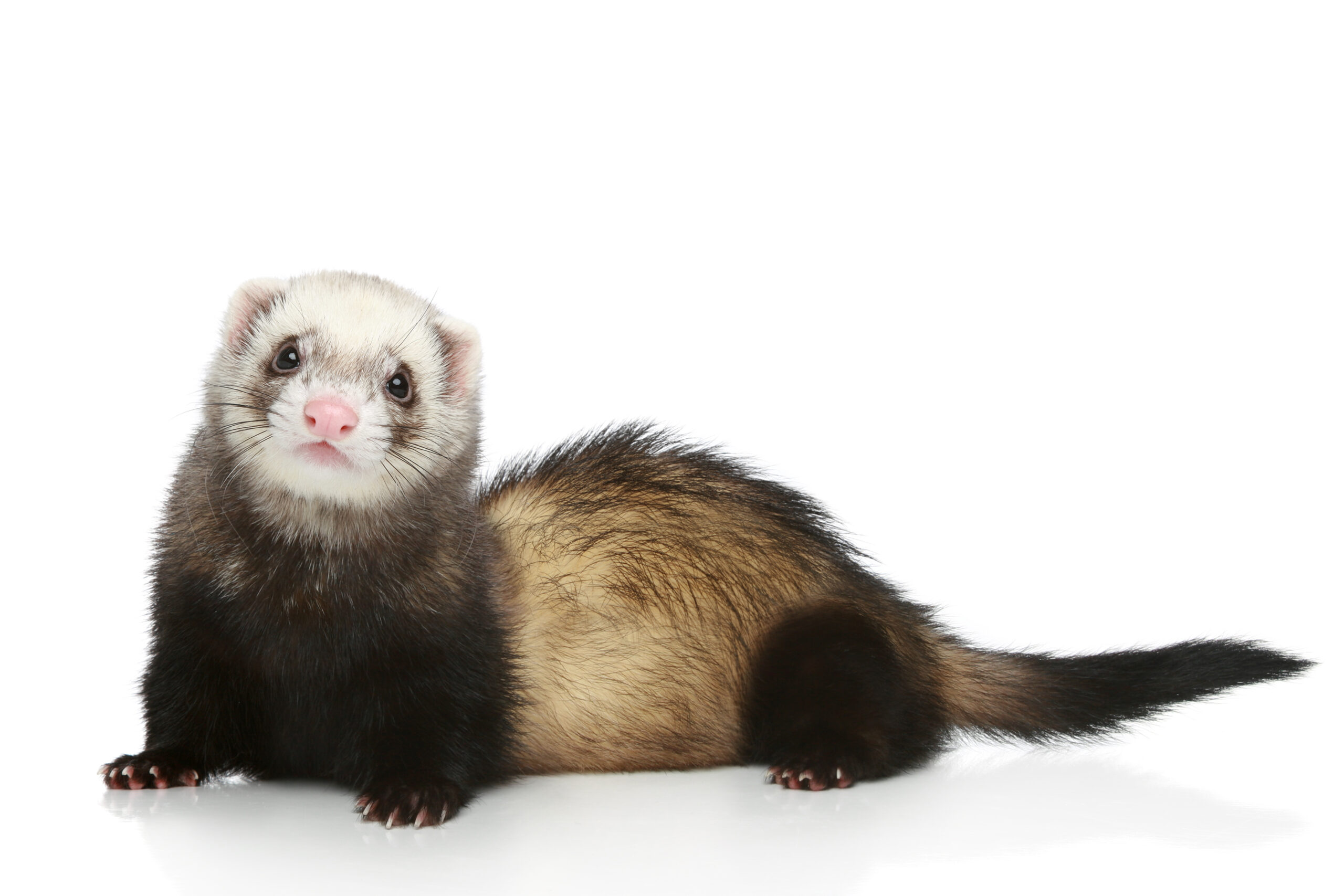
Ferrets
The ferret bladder is relatively small and thin walled and easily distensible. The male (hob) has a baculum that is J-shaped.(1) This runs from near the tip of the penis to caudal to the ventral rim of the pelvis. The urethral orifice opens on the ventral surface of the glans penis and the preputial opening is caudal to the umbilicus. Additionally, the prostate is fusiform in shape and surrounds the proximal urethra. Prostatic disease can occur as a sequela to hyperandrogenism associated with adrenal disease and mimic the presentation of urolithiasis. These anatomic variations result in a male ferret predilection for obstructive urolithiasis and may make catheterization challenging. The urethra in the female opens into the vaginal vestibule.
In ferrets, the range for creatinine is small and the total creatinine is lower compared to other species. Therefore, small elevations in creatinine may be significant in ferrets. Given their carnivorous diet, the urine pH is normally acidic.
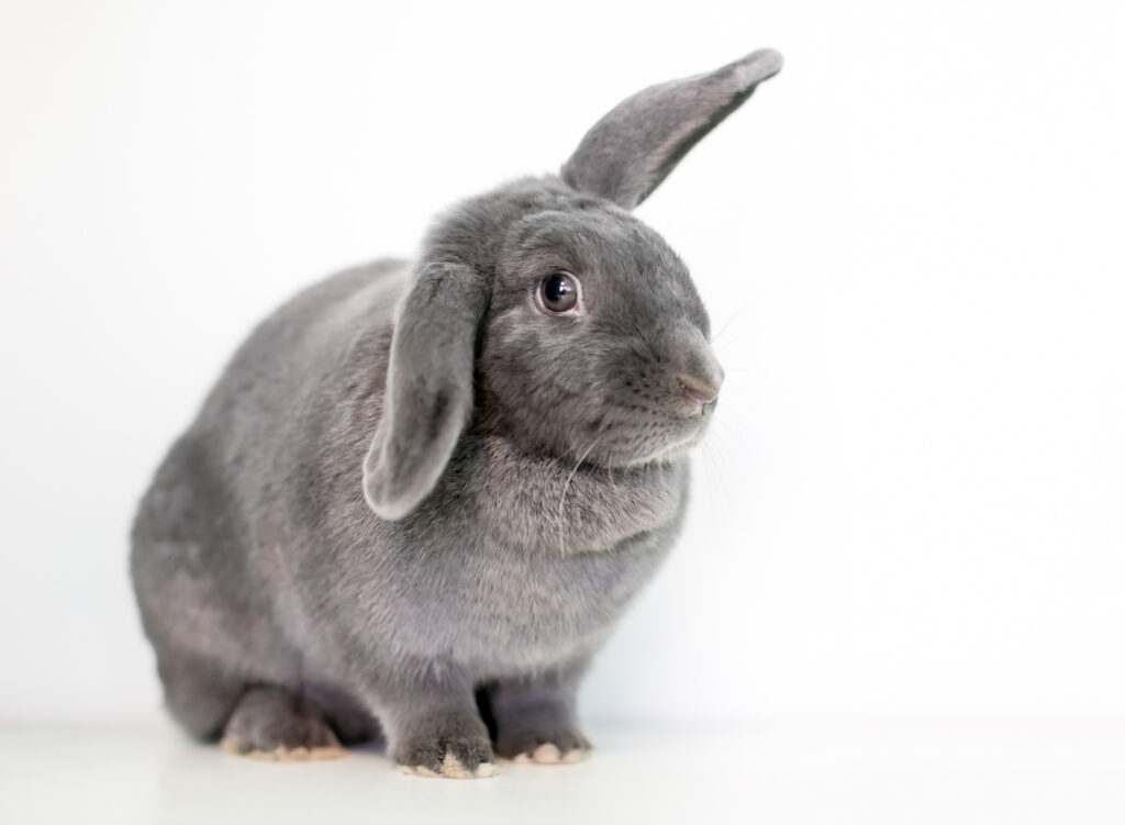
Rabbits
The bladder normally lies within the caudal abdomen to pelvic canal but is very distensible. There is no baculum in the male. The female urethra exits within the vaginal vestibule. Pigment from endogenously synthesized compounds or ingested plants are commonly excreted in the urine resulting in a yellow to red coloration. The urine is normally cloudy in color due to ammonium magnesium phosphate and calcium carbonate precipitates. Rabbits have a unique calcium metabolism compared to other mammals. Serum calcium is directly related to dietary calcium and intestinal absorption is independent of vitamin D levels. The urine is the main route of calcium excretion.(2,3) Rabbits may have less carbonic anhydrase within the thick ascending loop of renal tubules. However literature supporting this is sparse.(4,5) Rabbits have high water intake and urine output with daily fluid requirements of approximately 100 ml/kg/day.(6)
Guinea pigs
Males have numerous accessory sex glands including large vesicular glands in which stones can become lodged.(7) The penis is S-shaped and males have a baculum. There is intromittent sac containing two horny styles located caudoventral to the urethral opening at the tip of the glans penis. The female urethra is readily distensible, and the urinary papilla is located outside of the vagina, like most rodents. Females have a vaginal membrane. Guinea pigs also have a high urinary excretion of calcium.
Chinchillas
Male chinchillas possess a baculum. Males also have well developed accessory sex glands and produce a post-mating plug. Chinchillas can produce a wide range of urine specific gravities, but are often concentrated.(8,9) Chinchillas excrete the majority of their calcium in the feces, not urine.(10)
UROLITHIASIS BY SPECIES
Ferrets
Historically sterile struvite uroliths were the most common urolith type in the United States. In a study evaluating urolithiasis from the Minnesota Urolith Center from 1981-2007, 67% of the stones submitted were struvite.(11) However the prevalence of this stone type has shifted over time. In a more recent retrospective study (2010-2018), the most common stone type in ferrets in North America is now cystine, with 92.6% of submissions being primarily cystine in composition.(12) This is in contrast to uroliths in other countries with cystine urolithiasis making up only 26.8% of the submissions from Europe and Asia.(12) In this study, the odds of submission of a cystine urolith decreased with age with the median age of ferrets with cystine urolithiasis being 2 years of age and those with other stone types, 4 years of age.
Overall male ferrets appear to present more often with urolithiasis, likely due to their anatomical characteristics that predispose them to urinary obstructions.(11,12) Urinary pH may influence the type of stone that develops as well. Diets higher in plant proteins may produce more alkaline urine which favors the formation of struvite stones. While the naturally more acidic pH of ferret urine makes cystine insoluble.(1) In a retrospective case control study of ferrets with cystine urolithiasis, 94% of the cases with cystine stones received a grain-free diet.(13) While dietary management or prevention of this disease may be challenging, emphasis should be put on a high quality animal protein diet. Cystinuria is associated with an inherited genetic mutation in humans, dogs and cats.(14) Ferrets in the United States have minimal genetic diversity, and a similar underlying genetic mechanism likely contributes to this disease process.(15,16) Research is ongoing in this subject.
Rabbits
The most common urolith in rabbits is calcium carbonate. Other stone types include ammonium magnesium phosphate and calcium oxalate. Rare reports include silica, struvite and a single cases of calcium sulfate dihydrate from a rabbit eating gypsum based plaster.(17) The prevalence of urolithiasis in pet rabbit populations is unknown but estimated to be 2-10%.(18)
Most stones are located within the bladder, however urethroliths, ureteroliths and renoliths also occur. Rabbits can also develop uroliths within the vaginal vestibule secondary to urine pooling or uroliths that were urinated into the vestibule.(19)
In addition to true urolithiasis, rabbits may develop micro urinary calculi (MUC) which is more commonly referred to as bladder “sludge.” This is differentiated from normal calciuria as it precipitates to form microscopic calculi within the bladder. This appears to be a relatively common condition in rabbits.
Predisposing factors for naturally occurring urolithiasis are less well described in rabbits than in ferrets. Things like obesity, lack of voiding, infection and urinary nidus may universally predispose animals to stone formation. Ureteral obstruction itself can predispose rabbits to nephrolithiasis of the contralateral kidney.(20,21) Certain biochemical changes associated with urolithiasis in rabbits include increasing plasma calcium and sodium, likely due to their high calcium absorption and excretion.(18) A study evaluated the effect of dietary calcium on urolithiasis in rabbits fed diets of varying calcium concentrations for a total of 25 weeks. Rabbits fed primarily alfalfa, a high calcium food item, had more urinary sediment on ultrasound, but none of the rabbits in this study developed urolithiasis or tissue mineralization.(22) While diet is often blamed as a cause of urolithiasis in rabbits, calcium should not be excluded from the diet as it is a necessary nutrient. Instead, priority should be placed on increasing water intake and limiting dietary items with excessive calcium levels.
Check back next week for Part 2 HERE to learn more about guinea pigs and chinchillas, along with treatment of urolithiasis!
References for Part 1 and 2:
1. Di Girolamo N, Huynh M. Ch. 4 Disorders of the Urinary and Reproductive Systems in Ferrets In: Quesenberry K,Mans C, eds. Ferrets, Rabbits, and Rodents: Clinical Medicine and Surgery 4th eds. Philadelphia: Elsevier, 2020;39-55.
2. Eckermann-Ross C. Hormonal Regulation and Calcium Metabolism in the Rabbit. Vet Clin North Am Exot Anim Pract 2008;11:139-152.
3. Cheeke P, Amberg J. Comparative calcium excretion by rats and rabbits. Journal of animal science 1973;37:450-454.
4. Donnelly TM, Vella D. Ch. 11 Basic Anatomy, Physiology, and Husbandry of Rabbits In: Quesenberry KE, Orcutt CJ, Mans C, et al., eds. Ferrets, Rabbits, and Rodents (Fourth Edition). Philadelphia: W.B. Saunders, 2020;131-149.
5. Di Girolamo N, Selleri P. Ch 16. Disorders of the Urinary and Reproductive Systems In: Quesenberry KE, Orcutt CJ, Mans C, et al., eds. Ferrets, Rabbits, and Rodents (Fourth Edition). Philadelphia: W.B Saunders, 2020;201-219.
6. Brandao J, Graham J, Quesenberry K. Ch 12. Basic Approach to Veterinary Care of Rabbits In: Katherine Q, Christoph M, Connie O, et al., eds. Ferrets, Rabbits, and Rodents : Clinical Medicine and Surgery (4th edition). Philadelphia: W. B. Saunders, 2020;150-161.
7. Pignon C, Mayer J. Ch 21. Guinea Pigs In: Quesenberry KE, Orcutt CJ, Mans C, et al., eds. Ferrets, Rabbits, and Rodents (Fourth Edition). Philadelphia: W.B. Saunders, 2020;270-297.
8. Doss GA, Mans C, Houseright RA, et al. Urinalysis in chinchillas (Chinchilla lanigera). Journal of the American Veterinary Medical Association 2016;248:901-907.
9. Alworth LC, Harvey SB. Chapter 39 – Anatomy, Physiology, and Behavior In: Suckow MA, Stevens KA,Wilson RP, eds. The Laboratory Rabbit, Guinea Pig, Hamster, and Other Rodents. Boston: Academic Press, 2012;955-966.
10. Martel-Arquette A, Mans C. Urolithiasis in chinchillas: 15 cases (2007 to 2011). J Small Anim Pract 2016;57:260-264.
11. Nwaokorie EE, Osborne CA, Lulich JP, et al. Epidemiology of struvite uroliths in ferrets: 272 cases (1981–2007). Journal of the American Veterinary Medical Association 2011;239:1319-1324.
12. Hanak EB, Di Girolamo N, DeSilva U, et al. Variation in mineral types of uroliths from ferrets (Mustela putorius furo) submitted for analysis in North America, Europe, or Asia over an 8-year period. Journal of the American Veterinary Medical Association 2021;259:757-763.
13. Lamglait B, Brieger A, Rainville M-P, et al. Retrospective case control study of pet ferrets with cystine urolithiasis in Quebec, Canada: epidemiological and clinical features. Journal of veterinary medicine and surgery 2021;5.
14. Kovaříková S, Maršálek P, Vrbová K. Cystinuria in Dogs and Cats: What Do We Know after Almost 200 Years? Animals 2021;11:2437.
15. Gustafson KD, Hawkins MG, Drazenovich TL, et al. Founder events, isolation, and inbreeding: Intercontinental genetic structure of the domestic ferret. Evolutionary Applications 2018;11:694-704.
16. Stockman J, Malka S, Lofgren N, et al. Cystine and amino acid concentrations in the urine of pet ferrets (Mustela putorius furo). Journal of Exotic Pet Medicine 2023.
17. Kucera J, Koristkova T, Gottwaldova B, et al. Calcium sulfate dihydrate urolithiasis in a pet rabbit. Journal of the American Veterinary Medical Association 2017;250:534-537.
18. Wong A, Gardhouse S, Rooney T, et al. Associations between biochemical parameters and referral centre in pet rabbits with urolithiasis. Journal of Small Animal Practice 2021;62:554-561.
19. Tarbert DK, Matos Rd. Endoscopic Removal of a Vaginal Calculus in a Domestic Rabbit (Oryctolagus cuniculus). Journal of Exotic Pet Medicine 2016;25:253-260.
20. Itatani H, Yoshioka T, Namiki M, et al. Experimental model of calcium-containing renal stone formation in a rabbit. Investigative Urology 1979;17:234-240.
21. Eddy AA, Falk RJ, Sibley RK, et al. Subtotal nephrectomy in the rabbit: A model of chronic hypercalcemia, nephrolithiasis, and obstructive nephropathy. The Journal of Laboratory and Clinical Medicine 1986;107:508-516.
22. Clauss M, Burger B, Liesegang A, et al. Influence of diet on calcium metabolism, tissue calcification and urinary sludge in rabbits (Oryctolagus cuniculus). Journal of animal physiology and animal nutrition 2012;96:798-807.
23. Hawkins MG, Ruby AL, Drazenovich TL, et al. Composition and characteristics of urinary calculi from guinea pigs. Journal of the American Veterinary Medical Association 2009;234:214-220.
24. Parkinson LAB, Hausmann JC, Hardie RJ, et al. Urethral diverticulum and urolithiasis in a female guinea pig (Cavia porcellus). J Am Vet Med Assoc 2017;251:1313-1317.
25. Edell AS, Vella DG, Sheen JC, et al. Retrospective analysis of risk factors, clinical features, and prognostic indicators for urolithiasis in guinea pigs: 158 cases (2009–2019). Journal of the American Veterinary Medical Association 2022;1:1-6.
26. Gallego M. Case report of a satin guinea pig with fibrous osteodystrophy that resembles human pseudohypoparathyroidism. Case Reports in Veterinary Medicine 2017;2017.
27. Karam A, Mjaess G, Younes H, et al. Increase in urolithiasis prevalence due to vitamins C and D supplementation during the COVID-19 pandemic. Journal of Public Health 2022;44:e625-e626.
28. Singh P, Kiran R, Pendse A, et al. Ascorbic acid is an abettor in calcium urolithiasis: an experimental study. Scanning microscopy 1993;7:28.
29. Higbie CT, DiGeronimo PM, Bennett RA, et al. Semen-Matrix Calculi in a Juvenile Chinchilla (Chinchilla lanigera). Journal of Exotic Pet Medicine 2019;28:69-75.
30. Bartges JW, Callens AJ. Urolithiasis. The Veterinary clinics of North America Small animal practice 2015;45:747-768.
31. Huynh M. Permanent Implantable Medical Devices in Exotic Pet Medicine. Veterinary Clinics of North America: Exotic Animal Practice 2019;22:521-538.
32. Martorell J, Bailon D, Majó N, et al. Lateral approach to nephrotomy in the management of unilateral renal calculi in a rabbit (Oryctolagus cuniculus). Journal of the American Veterinary Medical Association 2012;240:863-868.
33. Rembeaux H, Langlois I, Burdick S, et al. Placement of ureteral stents in three rabbits for the treatment of obstructive ureterolithiasis. Journal of Small Animal Practice 2021;62:489-495.
34. Huynh M. Permanent Implantable Medical Devices in Exotic Pet Medicine. Veterinary Clinics: Exotic Animal Practice 2019;22:521-538.
35. Wenger S, Hatt J-M. Transurethral Cystoscopy and Endoscopic Urolith Removal in Female Guinea Pigs (Cavia porcellus). Veterinary Clinics of North America: Exotic Animal Practice 2015;18:359-367.
36. Branquart M, Langlois I, Vachon C, et al. Removal of lower urinary tract stones by percutaneous cystolithotomy in domestic male ferrets (Mustela putorius): 4 cases (2017–2020). Journal of Exotic Pet Medicine 2023;45:38-44.
37. Coutant T, Dunn M, Langlois I, et al. CYSTOSCOPIC-GUIDED LITHOTRIPSY FOR THE REMOVAL OF A URETHRAL STONE IN A GUINEA PIG. Journal of Exotic Pet Medicine 2019;28:111-114.
38. Maxwell AD, Kim GW, Furrow E, et al. Development of a Burst Wave Lithotripsy System for Noninvasive Fragmentation of Ureteroliths in Pet Cats. 2022.
39. Azevedo S, O’Malley B, Greene C, et al. Lower Urinary Tract Diseases in Guinea Pigs: A 14-Year Retrospective Study (2004–2018). Animals 2023;13:112.
40. Tschudin A, Clauss M, Codron D, et al. Water intake in domestic rabbits (Oryctolagus cuniculus) from open dishes and nipple drinkers under different water and feeding regimes. Journal of animal physiology and animal nutrition 2011;95:499-511.
41. Hagen K, Clauss M, Hatt JM. Drinking preferences in chinchillas (C hinchilla laniger), degus (O ctodon degu) and guinea pigs (C avia porcellus). Journal of animal physiology and animal nutrition 2014;98:942-947.


Only VETgirl members can leave comments. Sign In or Join VETgirl now!