


September 2023
By Dr. Garret Pachtinger, VMD, DACVECC, Director of Operations / Co-Founder, VETgirl
Cardiac Emergencies in the ER: Part 2 with Dr. Garret Pachtinger
In this VETgirl online veterinary CE blog, Dr. Garret Pachtinger, VMD, DACVECC reviews what you need to know about cardiac emergencies. If you haven’t already read Part 1 of this two-part blog “Cardiac Emergencies in the ER: Part 1,” make sure to read more about congestive heart failure, arrhythmias and more HERE.
Cardiopulmonary arrest / CPR
Cardiopulmonary arrest is associated with a poor prognosis and has not improved as technology and medicine have advanced. Moreover, most of the protocols previously used in veterinary medicine were extrapolated from human guidelines (American Heart Association). The Reassessment Campaign on Veterinary Resuscitation (RECOVER) recently completed a systematic review of the literature relevant to veterinary CPR and developed the first evidence-based, consensus CPR guidelines for small animals. RECOVER initiative is one of the first in veterinary medicine to evaluate CPR to create guidelines for CPR.
Basic Life Support (BLS)
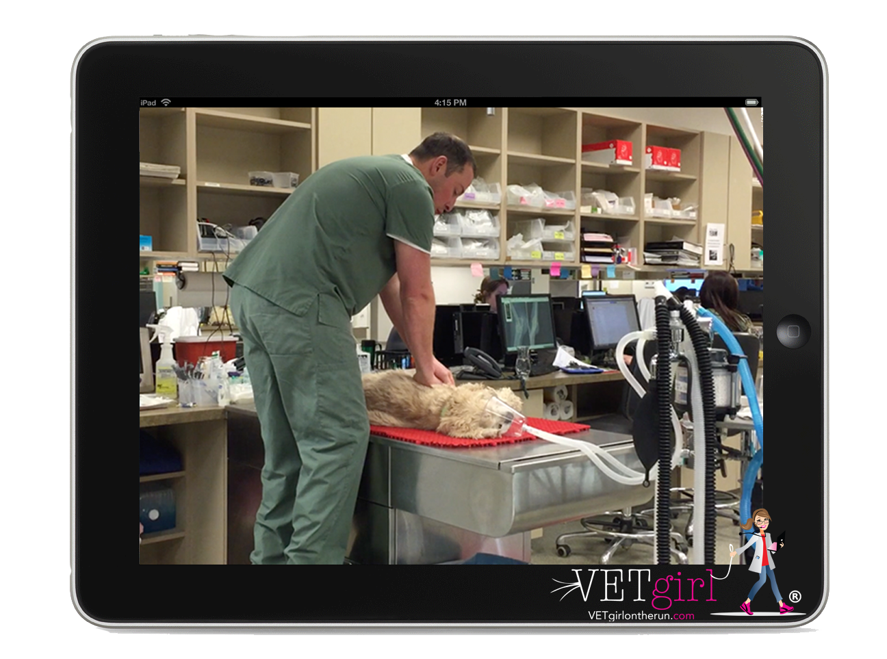
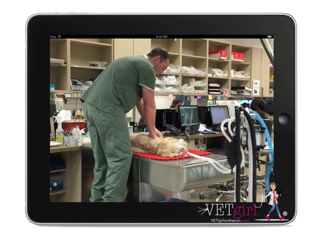
Basic Life Support (BLS) should be initiated as quickly as possible following diagnosis of cardiopulmonary arrest (CPA) including chest compressions, securing a patent airway, and ventilation.
Circulation: Chest Compressions
Patients with CPA have no forward blood flow out of the heart and therefore with a lack delivery of oxygen to the tissues and removal of waste products from the tissues, increasing the risk for hypoxemia, ischemic organ injury, and reperfusion injury if there is reinstitution of tissue blood flow. The goals of chest compressions are to provide pulmonary blood flow for oxygen uptake and waste elimination, and tissue perfusion for oxygen delivery to restore cellular metabolic activity.
There are 2 theories when initiating closed chest CPR.
“Cardiac Pump Theory”
This is most often considered for cats, small dogs, and keel chested larger dogs (such as Greyhounds, Dobermans). In this principle, the heart is considered the main element as the heart is Compressed between ribs. During this process, there is manual opening of the pulmonic and aortic valves leading to the forceful delivery of blood from the ventricles through the pulmonic and aortic valves. Retrograde flow is prevented via the closing of both AV valves followed by artificial diastole (rest and recoil) which refills the ventricles .
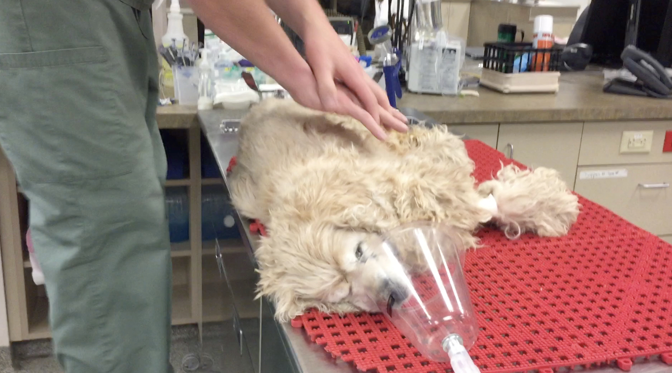
- Compressions occur directly over the heart (ventral 1/3 thorax over 4th-6th intercostals) with patient in lateral recumbency
- Rate: 100–120/minute
- Continue 10 breath per minute ventilation rate throughout the compression cycle
- Allow adequate recoil of the chest between compressions
Thoracic Pump Theory
This is most often considered for medium, large and giant round chested breeds (such as Labradors, Rottweilers). In this principle, the heart is considered a secondary element not primary. The goal is to have the forceful compressions change the intrathoracic pressure, resulting in opening and closing of the heart valves and movement of blood flow. Retrograde blood flow is prevented by the increase in intrathoracic pressure collapsing the thoracic vasculature
Important principles with this theory:
- Compressions take place at the widest part of the thorax, generally between the 7th-8th intercostal spaces
- Compressions must depress the thorax by 1/3
- Rate 100–120/minute
- Continue 10 breath per minute ventilation rate throughout the compression cycle
- Compression to relaxation ratio of 1:1 is ideal
- Allow adequate recoil of the chest between compressions
While most patients will benefit from CPR performed in lateral recumbency, broad chested dogs such as bulldogs may benefit from CPR performed in dorsal recumbency, similar to humans. When performing CPR in dorsal recumbency, compressions should depress the sternum 1 ½ to 2 inches.
Chest compressions should be delivered without interruption in cycles of 2 minutes. A new compressor begin chest compressions after each cycle to decrease the likelihood of fatigue blunting the effectiveness of CPR> Interruption in chest compressions should minimized to decrease the likelihood of a drop in coronary perfusion pressure (CPP). CPP is a critical determinant of myocardial blood flow and the likelihood of return of spontaneous circulation (ROSC).
Airway and Breathing: Ventilation
Although a delay in chest compressions should be minimized if at all possible, during CPR the patient should be intubated for airway control, oxygenation, and ventilation as soon as possible. Although not conventional for most veterinarians, intubation can be performed in lateral recumbency to minimize the need for cessation of chest compressions during endotracheal tube placement. An inspiratory time of approximately 1 second is recommended.
If an endotracheal tube is not readily available, mouth to snout ventilation is an alternative to improve oxygenation and waste product (CO2) removal. To perform mouth to snout ventilation, the patient’s mouth is held closed firmly with one hand and the neck is extended to align the snout with the spine. This allows opening of the airway to achieve the best oxygenation and ventilation The clinician then places their mouth over the patients nares and blows firmly into the nares to inflate the chest.
Again, chest compressions should not be delayed during intubation or ventilation by the mouth to snout technique, rather performed simultaneously during ventilation. Intubated patients should be ventilated at a rate of 10 breaths per minute with an inspiratory time of approximately 1 second. The tidal volume can be assessed using a spirometer, with the goal of approximately 10 ml/kg for each breath. Hyperventilation should be avoided as this may lead to a low CO2 and cerebral vasoconstriction, ultimately decreased perfusion.
Advanced Life Support (ALS)
Once BLS has been initiated, the CPR team should initiate Advanced Life Support (ALS). ALS focuses on drug therapy and electrical defibrillation.
Depending on the arrest rhythm, drug therapy may include the use of vasopressors, parasympatholytics, and/or anti-arrhythmics, reversal agents, intravenous fluids, and alkalinizing drugs. For this reason, placement of a peripheral or central intravenous or intraosseous catheter is recommended.
Vasopressors
Vasopressors are recommended to increase peripheral vasoconstriction and improve cardiac output as even the most forceful chest compressions will not mimic the cardiac function of a healthy patient. It is up to the clinician to then consider medications to improve perfusion to the vital organs including the heart, lungs, and brain,
- Epinephrine causes peripheral vasoconstriction via stimulation of α1adrenergic receptors as well as β1 and β2
- Low doses (0.01 mg/kg) is recommended initially. Following prolonged CPR, high dose administration may be considered (0.1 mg/kg)
- While recommended via the intravenous or intraosseous route, epinephrine may also be administered via ET tube (double the dose – 0.02 mg/kg low dose; 0.2 mg/kg high dose) via a long catheter through the endotracheal tube and diluting 1:1 with isotonic (0.9%) saline.
- Vasopressin exerts its vasoconstrictive effects via activation of peripheral V1 receptors
- This can be used in combination or in place of epinephrine at a dose of 0.8 U/kg IV/IO.
- Potential benefits of vasopressin include efficacy in acidic environments in which α1receptors may become unresponsive to epinephrine
- Vasopressin lacks β1effects which may cause increased myocardial oxygen consumption and worsened myocardial ischemia upon ROSC.
- Vasopressin may also be administered via endotracheal tube using the technique described for epinephrine above.
Anticholinergics
Atropine is the common anti-cholinergic considered in CPR and is a parasympatholytic medication. Dosed at 0.04 mg/kg IV/IO is considered during CPR in dogs and cats when the cause is considered a result of asystole or PEA associated with increased vagal tone. Atropine may also be administered via ET tube (0.08 mg/kg).
Anti-arrhythmics
Ventricular fibrillation or pulseless ventricular tachycardia (VT) should be treated as early as possible with electrical defibrillation. If electrical defibrillation is not corrective, drug therapy may be warranted. Amiodarone, dosed at 2.5–5 mg/kg IV/IO is considered. Alternatively, if not available lidocaine 2 mg/kg slow IV/IO is an alternative. Side effects of amiodarone includes anaphylactic reactions, hypotension, peripheral vasodilation, wheals, or hives. Treatment with diphenhydramine (2 mg/kg IM) and/or anti-inflammatory corticosteroids (0.1 mg/kg dexamethasone sodium phosphate IV) is recommended if these signs are seen following ROSC.
Reversal Agents
Reversal agents are considered when reversible anesthetic drugs were recently administered. Naloxone (0.01 mg/kg IV/IO) may be used to reverse opioids, flumazenil (0.01 mg/kg IV/IO) for benzodiazepines, and atipamezole (0.1 mg/kg IV/IO) or yohimbine (0.1 mg/kg IV/IO) for α2 agonists.
Intravenous (IV) Fluids
Intravenous fluids in euvolemic or hypervolemic patients is not recommended during CPR as it will increase right atrial pressure resulting in decreased perfusion of the brain and heart. Conversely, hypovolemic patients, IV fluids are recommended to restore adequate circulating volume and increase the efficacy of chest compressions with improved perfusion.
Corticosteroids
The use of corticosteroids during CPR in dogs and cats is not recommended. There is not a body of evidence which shows significant benefit from the use of steroids during CPR. Moreover, side effects of steroid use include gastric ulceration, suppression of the immune system, and reduced prostaglandin production in the kidney.
Alkalinization Therapy
In patients with prolonged CPA (greater than 10–15 minutes), alkalinization therapy is considered. Sodium bicarbonate dosed at 1 mEq/kg (diluted IV) may be considered. The rationale for this therapy is that prolonged CPA results in severe acidemia (lactate acid, uremic acids, and venous respiratory acidosis) due to poor perfusion and accumulation of CO2. The acidemia ultimately results in vasodilation and inhibition of normal enzymatic and metabolic activity.
Electrical Defibrillation
Electrical defibrillation is considered in patients with ventricular fibrillation (VF). Defibrillators may be either monophasic (delivering a current in one direction across the paddles) or biphasic (delivering a current in one direction, the reversing and delivering a current in the opposite direction).
- The use of biphasic defibrillators is recommended because a lower current is required to successfully defibrillate the patient, decreasing the likelihood of myocardial injury.
- Monophasic defibrillators are used at an initial dose of 4–6 J/kg while biphasic defibrillation should initially used at 2–4 J/kg. If the first defibrillation is unsuccessful, a second attempt can be tried by increasing the dose by 50%.
Following defibrillation, chest compressions should be resumed immediately. A complete 2-minute CPR cycle should be performed before reassessing the ECG and determining if the patient requires additional defibrillation for continued ventricular tachycardia.
Open Chest CPR
As compared to closed-chest CPR, open-chest CPR results in improved output. Importantly, open-chest CPR is more invasive, expensive and requires significant post ROSC planning. Indications for open chest CPR include pericardial disease, pleural space disease, and thoracic wall defects such as numerous rib fractures or a flail chest. If the patient is under general anesthesia for an abdominal procedure, direct cardiac massage can be performed with an incision into the diaphragm. Finally, giant breed and very large chested dogs may not respond to closed chest CPR and for these patients, open chest CPR should be considered.

Additional monitoring
Commonly used monitoring devices for CPR include pulse oximetry, direct and indirect blood pressure monitors, continues electrocardiogram (ECG) and end tidal CO2 monitor (ETCO2).
Electrocardiogram (ECG)
ECG monitoring during CPR is used to diagnose the arrest rhythm
- Asystole
- Pulseless electrical activity (PEA)
- Ventricular fibrillation (VF).
The ECG is also used to determine if there is a change in the cardiac rhythm during and following therapy.
End-Tidal Carbon Dioxide
ETCO2 during CPR has several indications. One indication is to determine if endotracheal intubation was successful. If the endotracheal tube was placed mistakenly in the esophagus, there will be little to no reading. If there is measurable CO2 by ETCO2, this would be supportive of correct placement of the endotracheal tube. Importantly, if there is return of spontaneous circulation (ROSC), ETCO2 will rapidly increase due to the rapid increase in circulation. It is therefore used as an early indicator of ROSC during CPR.
Conclusion
When in doubt, the veterinarian must feel comfortable managing cardiac emergencies in the veterinary ER to help improve overall survival.
References (for both Part 1 and 2 of this blog):
- Andreka P, Frenneaux MP. Haemodynamics of cardiac arrest and resuscitation.Curr Opin Crit Care. 2006;12(3):198–203.
- Berg RA, Otto CW, Kern KB,et al. A randomized, blinded trial of high-dose epinephrine versus standard-dose epinephrine in a swine model of pediatric asphyxial cardiac arrest. Crit Care Med. 1996;24(10):1695–1700.
- Berg RA, Otto CW, Kern KB,et al. High-dose epinephrine results in greater early mortality after resuscitation from prolonged cardiac arrest in pigs: a prospective, randomized study.Crit Care Med. 1994;22(2):282–290.
- Buckley GJ, Rozanski EA, Rush JE. Randomized, blinded comparison of epinephrine and vasopressin for treatment of naturally occurring cardiopulmonary arrest in dogs.J Vet Intern Med. 2011;25(6):1334–1340.
- Fletcher DJ, Boller M, Brainard BM,et al. RECOVER evidence and knowledge gap analysis on veterinary CPR. Part 7: Clinical guidelines. J Vet Emerg Crit Care (San Antonio). 2012;22(Suppl 1):S102–131.
- Hopper K, Epstein SE, Fletcher DJ,et al. RECOVER evidence and knowledge gap analysis on veterinary CPR. Part 3: Basic life support. J Vet Emerg Crit Care (San Antonio). 2012;22(Suppl 1):S26–43.
- Iwami T, Kitamura T, Kawamura T,et al. Chest compression-only cardiopulmonary resuscitation for out-of-hospital cardiac arrest with public-access defibrillation: a nationwide cohort study. Circulation. 2012;126(24):2844–2851
- Kern KB, Hilwig R, Ewy GA. Retrograde coronary blood flow during cardiopulmonary resuscitation in swine: intracoronary Doppler evaluation.Am Heart J. 1994;128(3):490–499.
- Lindner K, Haak T, Keller A,et al. Release of endogenous vasopressors during and after cardiopulmonary resuscitation. Heart. 1996;75(2):145–150.
- Lindner KH, Dirks B, Strohmenger HU,et al. Randomised comparison of epinephrine and vasopressin in patients with out-of-hospital ventricular fibrillation. Lancet. 1997;349(9051):535–537.
- Mentzelopoulos SD, Zakynthinos SG, Siempos I,et al. Vasopressin for cardiac arrest: meta-analysis of randomized controlled trials. Resuscitation. 2012;83(1):32–39.
- Paradis NA, Martin GB, Rivers EP,et al. Coronary perfusion pressure and the return of spontaneous circulation in human cardiopulmonary resuscitation. J Am Med Assoc. 1990;263(8):1106–1113
- Rozanski EA, Rush JE, Buckley GJ,et al. RECOVER evidence and knowledge gap analysis on veterinary CPR. Part 4: Advanced life support. J Vet Emerg Crit Care (San Antonio). 28.
- Stiell IG, Hébert PC, Wells GA,et al. Vasopressin versus epinephrine for inhospital cardiac arrest: a randomised controlled trial. Lancet. 2001;358(9276):105–109.
- Wik L, Naess PA, Ilebekk A,et al. Effects of various degrees of compression and active decompression on haemodynamics, end-tidal CO2, and ventilation during cardiopulmonary resuscitation of pigs. Resuscitation. 1996;31(1):45–57
- Borgeat K, Wright J, Garrod O, et al. Arterial thromboembolism in 250 cats in general practice: 2004–2012. J Vet Intern Med. 2012;28:102–108.
- Smith, F.W.K., Keene, B.W.: Left Sided Congestive Heart Failure. In The Five Minute Veterinary Consult, 3rd(eds. L. Tilley and F.W.K. Smith) Baltimore, Lippincott, Williams & Wilkins, 2004.
- Tilley, L.P., and Goodwin, J.: Manual of Canine & Feline Cardiology, 3rdPhiladelphia, W.B. Saunders, 2000.
- Norsworthy, G., Crystal, M., Fooshee, S., Tilley, L.P.: The Feline Patient.Baltimore, Lippincott, Williams & Wilkins, 1998.
Only VETgirl members can leave comments. Sign In or Join VETgirl now!


If ROSC is achieved is there serial labs that could also be used to assess the patient along with the monitoring of ecg, blood pressure, and ETCo2? Possible checking blood gasses, blood glucose, and electrolytes would give any additional indications of a possible CPA event recurring.
Wish we had electrical defibrillators.
I have tried closed chest compression CPR with ventilation and drug therapy, but I would not feel comfortable trying open chest CPR
chest compression CPR with ventilation and drug therapy, but I would not feel comfortable trying open chest CPR
Good information to have on hand in case of emergency
Good emergency information. Thank you.